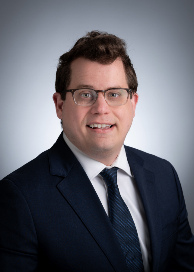
 Tylor Lewis, Ph.D.Tylor Lewis, Ph.D., assistant professor in the Department of Ophthalmology and Visual Sciences, has been named the latest recipient of the school's Featured Discovery award. This recognition celebrates notable faculty research contributions and highlights the impact of their scientific advancements.
His study, “Adam9-deficient retinal pigment epithelium pseudopods maintain photoreceptor outer segment renewal despite subretinal space expansion,” was published in the Journal of Clinical Investigation. The findings were also highlighted in an accompanying editor's commentary, which noted the work's potential to reshape understanding of how retinal cells maintain vision despite disease-related separation.
A common belief in visual science is that the light-sensitive photoreceptor cells and the retinal pigment epithelium (RPE) require close interaction in order to maintain visual health. The reason for this is that the RPE provides vital support to the photoreceptors. However, this isn’t always the case.
According to Lewis, certain clinical studies have observed that the separation of photoreceptors and RPE can sometimes have no negative effect on vision. The study aimed to understand what causes this outcome, as well as how to replicate it for the benefit of patients.
“We found that the RPE can form elongated structures known as ‘pseudopods’ to reach out and maintain contact with photoreceptors in order to support their function and health in disease,” said Lewis. “This work provides a biological foundation for our understanding of the slow progression of several vision diseases and provides an opportunity to promote this cellular process as an avenue to extend visual health in these patients.”
The Heersink communications team met with Lewis to gain insights into the study and help raise awareness about both the research and the Heersink School of Medicine.
What compelled you to pursue this research?
At the start of this project, my research was largely focused on the basic mechanisms underlying the function and maintenance of light-sensitive photoreceptor cells. For this project, I give a lot of credit to interacting with clinicians who treat patients with retinal degenerative diseases. After coming together and talking, we realized that the scientific perspective on how certain retinal cells interact did not fully explain the clinical outcomes of some retinal diseases. This pushed us to this line of research in order to better understand this apparent paradox.
When did you know you had an important discovery?
One of my favorite experimental techniques is electron microscopy. I remember sitting at the transmission electron microscope (TEM) and observing these elongated RPE structures for the very first time, thinking that this had to be the explanation. The main question in my mind at the time was whether or not these structures were actually functional. Once I discovered that they were functional and did indeed support photoreceptor function, I knew this would be an important research direction for years to come.
What was your most unexpected finding?
In patients with vitelliform macular dystrophy, a large separation between photoreceptors and the RPE can exist for years without any major disruptions in vision, until the sudden onset of significant vision disruption. After our initial discovery of the RPE pseudopods, I think the most unexpected result is that, in our model, the onset of retinal degeneration was associated with a reduction in the ability of the RPE to form these structures.
While unexpected, this finding was really exciting for two reasons. First, it provided a possible explanation for why photoreceptor cells eventually degenerate. Second, I saw it as an opportunity to explore ways in which we could try to promote this cellular process to potentially extend the lifespan of photoreceptors in these disease conditions.
What is the relevance of your research to human disease?
We believe that there are two major areas of relevance for human disease. First, this plasticity of the RPE to form pseudopods provides an explanation for the relatively slow progression of retinal diseases characterized by separation of these two cell types, such as Best disease and age-related macular degeneration (AMD) associated with subretinal drusenoid deposits. Second, we hope that by understanding the molecular mechanisms underlying this RPE plasticity, we can develop therapeutic strategies to promote pseudopod formation and prolong retinal health and function in these patients, who may otherwise ultimately go blind.
What made you come to UAB?
UAB has a long-standing history of strength in vision science research. Before coming here to start my own independent lab, I was continuously impressed by the interactions I had with UAB vision scientists, both within the Department of Ophthalmology and Visual Sciences (DOVS) in the Heersink School of Medicine, as well as university-wide scientists that are part of the Vision Science Research Center. The research support at UAB has been phenomenal, and I am incredibly excited to have joined the DOVS to help build on this history of excellence in vision science.