Case History:
A middle-age male patient with lytic sphenoid sinus lesion.
What is the diagnosis?
A. Myoepithelial carcinoma
B. Chondrosarcoma
C. Chondromyxoid fibroma
D. Pleomorphic adenoma
Correct Answer: C. Chondromyxoid fibroma
Discussion:
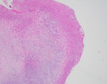
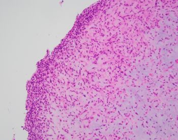
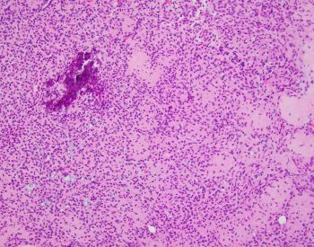
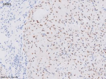
Chondromyxoid fibroma is a benign neoplasm that commonly involves the craniofacial (jaw and sinonasal) bones. Histologically, it is comprised of lobules of spindled and stellate cells with chondromyxoid background, arranged in a zonal architecture (i.e. hypercellular periphery and hypocellular center). Coarse calcifications can be seen. GRM1 expression by immunohistochemistry is a good surrogate marker for GRM1 gene recombination which is present in the majority of CMF cases.
Reference(s):
Purgina B, Baumhoer D, et al. Chondromyxoid fibroma. In: WHO Classification of Tumours Editorial Board. Head and neck tumours [Internet]. Lyon (France): International Agency for Research on Cancer; 2023 [cited 2024 Dec 19]. (WHO classification of tumours series, 5th ed.; vol. 9). Available from: https://tumourclassification.iarc.who.int/chapters/52/171.
Case contributed by: Melad Dababneh, MBBS, Assistant Professor, Anatomic Pathology